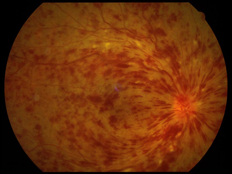
CRVO - Retinal hemorrhages all over with blood and thunder appearance
Central Serous Retinopathy - Blister of fluid at the macula - causes micropsiaMacular hole, differentiated from CRAO by absence of retinal pallor.
Add caption
Add caption
Add caption
Add caption
Add caption
Add caption
Add caption
Add caption
Best's Disease: Egg Yolk like appearance at the macula Headlight in the fog appearance in Toxoplasmosis Snow banking/Snowbank exudates in Pars Planitis
Fundus photographs of the patient before (A) and after (B) dark adaptation. The fundus appearance was characteristic of Oguchi disease with grayish-yellow discoloration that had normal appearance after 2–3 h of dark adaptation.- MIZUO phenomenon CMV Retinitis - Pizza Pie or Cottage Cheese and Tomato Ketchup retinopathy Corneal Endothelium seen by Specular Microscopy Cobblestone papillae in Vernal Conjunctivitis (spring catarrh) Horner Trantas Spots in vernal conunctivitis (spring catarrh) Amsler grid for monitoring macular pathology (ARMD, drusen etc) Pterygium
Cornea verticillata - Vortex keratopathy Band Keratopathy
Bitot's spots
Blepharitis
Chalazion
Hordeolum externum - stye - at base of hair follicle
Incision and Curettage of a chalazion with chalazion clamp also seen Sebaceous gland carcinoma - maybe mistaken as recurrent chalazion Acute dacryocystitis - treat with antibiotics; definitive treatment is DCR once the infections settles down.
Congenital dacryocystitis
Bowman's lacrimal probe and Nettleship punctum dilator Dacryoadenitis - S shaped ptosis Right orbital floor fracture - Tear drop sign - orbital contents prolapsing into maxillary sinus
Signs of orbital blow out fracture Lagophthalmos of left eye with exposure keratopathy of left inferior cornea
Corneal abrasion stained by fluorescein and viewed with cobalt blue light - epithelial defects stains underlying stroma with fluorescein